Advancements and Clinical Utility of Rigid Video Esophagoscope
The advent of the Rigid Esophagoscope has brought notable evolution in the field of esophageal diagnostics and interventions. In its first paragraph, it is apt to emphasize that unlike conventional rigid esophagoscopes, the rigid video variant integrates a telescope, light source, endo-camera, sheath, and an instrumentation channel into a compact unit. This “5-in-1” design allows for real-time visualisation, tissue sampling, foreign body removal and dilation procedures—all through a single rigid instrument—thus enhancing procedural efficiency and reducing the need for multiple adjuncts.
In this article, we will explore the history, design, clinical applications, procedural technique, advantages and limitations, safety concerns, comparisons with flexible endoscopy, and future directions of rigid video esophagoscopy.
Historical Context and Evolution
Rigid esophagoscopy has a long tradition in otolaryngology and surgical practice. For decades, rigid tubular metal scopes with optical lenses and illumination were used to inspect, biopsy, dilate, or extract foreign bodies from the esophagus. Over time, flexible fiberoptic and video endoscopes came to dominate because of their bendability and easier patient tolerance. Nevertheless, rigid tools continue to have a role, particularly when stable access, firm control, and wide working channels are needed.
Modern rigid video esophagoscopes combine the advantages of rigid systems (stability, larger instrumentation channel) with integrated imaging and lighting, making them more compact, cost-effective, and versatile, especially in settings with limited resources.
Structure and Design Features
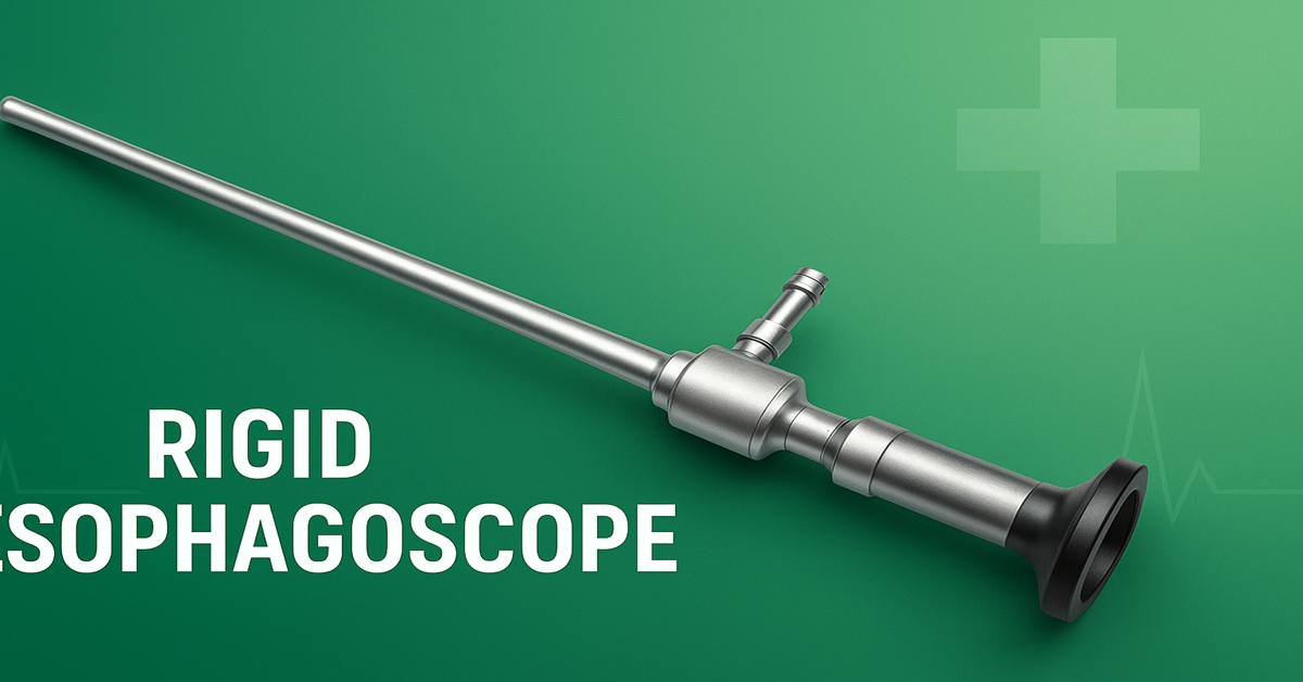
A typical modern rigid video esophagoscope is constructed of stainless steel, with a fixed obturator or tip made of a biocompatible material (such as Delrin) to mitigate trauma. Within its structure are embedded a miniaturized camera and LED light sources (often six LEDs) near the tip to illuminate the lumen. The video signal travels via fine wiring through the scope to an internal analog-to-digital converter, with output via a USB interface. This allows streaming, recording, and viewing on various display devices (e.g. desktops, laptops, tablets or smartphones).
The device also incorporates an instrumentation channel (commonly about 2.5 mm internal diameter) through which micro-forceps, biopsy tools, or retrieval devices can be passed. A side port may serve as a gas or air insufflation channel. These scopes are often designed with zero-degree vision and working lengths of 30–40 cm, and outer diameters approximately 8 mm, though pediatric variants may differ. The entire instrument is made sterilizable by methods such as ethylene oxide gas or formalin chamber immersion.
Compared to conventional rigid esophagoscopes that require external light pipes, camera adapters, and separate sheaths, the rigid video model’s integrated form reduces instrument bulk, setup time, and cost. Indeed, such devices may weigh only around 120 g, making them easier to handle during delicate maneuvers.
Clinical Indications and Applications
Rigid video esophagoscopes are applied across a spectrum of diagnostic and therapeutic tasks in the esophagus, especially in the upper (cervical) region. Key indications and applications include:
-
Foreign Body Removal
One of the oldest and most critical roles of rigid esophagoscopy is the safe removal of foreign bodies lodged in the esophagus. Rigid scopes allow better control, force application, and direct vision to grasp and extract coins, bones, or other ingested items. -
Detection and Biopsy of Lesions
Suspicious strictures, masses, or mucosal abnormalities may be visualized and biopsied via the instrumentation channel under direct vision. This is useful in diagnosing esophageal cancer, Barrett’s changes, or inflammatory lesions. -
Dilation of Strictures
In patients with esophageal narrowing (strictures), the rigid video esophagoscope can facilitate controlled dilation. A dilator or balloon may be passed through the instrument channel or advanced over the scope under visualization. -
Therapeutic Interventions
In selected cases, the rigid video esophagoscope supports superficial therapies such as injection, bleeding control, or limited mucosal resections, though its therapeutic reach is more limited compared to larger flexible platforms. -
Evaluation of Upper Esophagus / Hypopharynx Region
Because the rigid scope provides excellent straight-line access, it is often superior in the examination of the hypopharynx, upper esophageal sphincter, and cervical esophagus—areas where flexible scopes may face difficulty. -
Resource-Limited Settings
In environments where access to advanced flexible endoscopes or full video towers is limited, a rigid video esophagoscope offers a cost-effective alternative for basic esophageal examination and interventions.
However, rigid scopes are less effective for inspecting the distal (lower) third of the esophagus, where flexibility becomes essential.
Procedural Technique and Steps
The procedure typically unfolds as follows:
-
Preoperative Preparation
Patients must fast per anesthesia protocol. Medical history, medication review, and airway assessment are performed. Dental status is checked, as trauma to teeth is a risk. Consent is obtained. -
Anesthesia and Positioning
Rigid esophagoscopy is usually performed under general anesthesia with endotracheal intubation, to secure airway and suppress reflexes. The neck is extended using a head ring and shoulder roll so as to create a straight passage. The table may be rotated for surgeon ease. -
Scope Insertion
The scope, often with its obturator in place, is advanced through the oral cavity, taking care to protect lips and teeth (a dental guard may be used), and maintaining midline advancement. The bevel or tip is oriented posteriorly to enter the upper esophageal sphincter. Gentle pressure is applied to navigate resistance. -
Visualization and Intervention
Once in the esophagus, the video feed enables direct inspection. Through the instrumentation channel, biopsy forceps can be introduced, foreign bodies grasped and removed, dilation devices advanced, or bleeding controlled. After intervention, the region is re-inspected to check for residual injury or bleeding. -
Withdrawal and Final Checks
The scope is slowly withdrawn, observing for signs of mucosal tear or perforation. If any suspicious perforation is encountered, contrast studies like a barium swallow may be ordered. -
Postoperative Care
Patients are monitored until wakefulness and stable vital signs. Pain control, dietary instructions, and follow-up imaging or clinical checks are arranged. Any signs of fever, chest pain, mediastinal/emphysema signs, or bleeding prompt further evaluation.
Advantages of Rigid Video Esophagoscope
The rigid video esophagoscope offers several compelling benefits compared to traditional rigid scopes and even flexible endoscopes in certain scenarios:
-
Compact and Integrated Design
By integrating telescope, camera, lighting, sheath, and instrumentation in one unit, the instrument reduces the need for external cameras, light sources, or adapters, thereby simplifying setup and maintenance. -
Cost Efficiency
Because the design consolidates components, the device can be manufactured at a fraction of the cost of full-scale video endoscope systems. In some markets, such devices are advertised at a small fraction of conventional endoscopic systems. -
Good Optical Quality
With chip-on-tip technology and onboard LEDs, image clarity is maintained without loss due to intermediate fiberoptic or relay systems. -
Improved Control and Instrument Channel
The rigidity allows the surgeon to manipulate the scope with stability, and the instrumentation channel supports the passage of tools that may be more restricted in narrow flexible scopes. -
Accessible Technology in Low-Resource Settings
For settings lacking full endoscopy towers, the USB output and portability allow display with common computing devices, making it accessible and mobile. -
Faster Turnaround
Because fewer external components are needed, cleaning, calibration, and handling can be faster, which is advantageous for high-volume or emergency settings.
Limitations and Challenges
Despite its advantages, rigid video esophagoscopy also bears several limitations:
-
Limited Reach to Distal Esophagus
Rigid scopes are less effective for reaching far into the thoracic esophagus or gastroesophageal junction; flexible scopes are superior in that respect. -
Requirement of General Anesthesia
Unlike flexible endoscopy (which may be done under sedation), rigid scope procedures almost always require general anesthesia, with its attendant risks and logistical demands. -
Risk of Mucosal Injury and Perforation
The rigid nature increases the risk of rubbing, tearing, or perforation of the esophageal wall or surrounding tissues. Care must be taken in navigation and force application. -
Dental Trauma and Soft Tissue Injury
Teeth, lips, and oral structures may incur trauma if the scope is advanced without protection or caution. -
Limited Flexibility and Angulation
The rigid tube cannot negotiate curves or tortuous anatomy, so in patients with anatomical challenges (e.g. limited neck extension or trismus), access may be compromised. -
Sterilization and Mechanical Wear
The internal camera, wiring, tip, and optics demand careful handling during sterilization cycles. Repeated sterilization may degrade components over time. -
Smaller Working Channel
The instrumentation channel is often quite small (e.g. 2 mm cross-section), limiting the size and variety of tools that can be passed.
Safety Considerations and Risks
Performing rigid esophagoscopy demands meticulous technique and awareness of potential hazards:
-
Esophageal Perforation
Perhaps the gravest risk, perforation may lead to mediastinitis, infection, sepsis, or even death if not promptly managed. Vigilance for resistance, unexpected bleeding, or abnormal anatomy is crucial. -
Bleeding or Mucosal Tear
Manipulation or biopsy may cause bleeding or superficial mucosal injury. Hemostasis must be ensured before withdrawal. -
Anesthesia-related Complications
Risks associated with general anesthesia (respiratory events, cardiovascular instability) must be mitigated by proper monitoring and preoperative evaluation. -
Airway and Shared Space Hazards
Because the airway and esophagus share anatomical space, coordination between anesthesiologist and surgeon is essential to prevent tube displacement or airway compromise. -
Dental or Oral Injury
Propping open the mouth, guarding teeth, and using dental protection devices help minimize chip or fracture risk. -
Device Malfunction
Camera failure, lighting loss, or wiring breakage may impair procedure. Backup plans must exist. -
Delayed Complications
Postoperative fever, chest pain, subcutaneous emphysema, or dysphagia may signal complications requiring urgent assessment.
Surgeons must adopt a low threshold to abort the procedure or convert to open surgical management if complications or resistance is encountered.
Comparisons: Rigid Video vs Flexible Endoscopy
Flexible endoscopy (often via fiberoptic or video gastroscopes) is the predominant modality for esophageal evaluation today, owing to its bendability, patient comfort, and minimal sedation requirements. However, each modality has advantages and trade-offs:
-
Accessibility vs Reach
Flexible scopes can traverse the entire esophagus and beyond, while rigid scopes are more limited toward distal areas. -
Comfort and Sedation
Flexible scopes often require only sedation or topical anesthesia, avoiding full general anesthesia. Rigid scopes typically necessitate general anesthesia. -
Instrumentation Capability
Rigid scopes allow stronger manipulation and more stable insertion of tools; flexible scopes may have narrower working channels and less mechanical leverage. -
Vision and Imaging
Modern flexible video endoscopes offer high-definition imaging, zooming, and even narrow-band or advanced imaging modes. Rigid video scopes must maintain comparable optical quality to compete. -
Safety
While both carry risks, flexible scopes may lead to lower rates of perforation in expert hands; rigid scopes demand particularly careful technique. -
Cost and Infrastructure
High-end flexible endoscopic systems are expensive and require towers, processors, light sources, and maintenance. Rigid video scopes, especially USB-based ones, are comparatively more affordable and simpler to maintain.
In many cases, these modalities complement each other: rigid video esophagoscopy may be the tool of choice for foreign body extraction or in resource-constrained settings, whereas flexible endoscopy is preferred for routine diagnostics and deeper reaches.
Future Directions and Innovations
The field of rigid video esophagoscopy, though not novel, still holds room for innovation:
-
Miniaturization and Enhanced Optics
Further shrinking the camera, wiring and increasing LED brightness or using advanced CMOS chips could improve lumen illumination and allow smaller diameters. -
Enhanced Instrument Channels
Wider or dual channels may enable more robust therapeutic tools, suction, irrigation, or robotic instruments. -
Smart Imaging
Incorporation of real-time image processing, narrow-band imaging, or fluorescence could improve mucosal detection and diagnostic yield. -
Wireless or Fiberless Designs
Future designs might eliminate wired USB output in favor of wireless streaming, reducing tether constraints. -
Hybrid Rigid-Flexible Systems
A semi-rigid but articulating rigid video esophagoscope may permit better access while maintaining stability. -
Training and Simulation Models
Development of simulators for rigid video esophagoscopy will help clinicians safely gain experience before performing on patients. -
Cost Reduction and Dissemination
Wider adoption in low- and middle-income settings will depend on further cost optimization, robust design, and ease of sterilization and maintenance.
Summary and Conclusion
Rigid video esophagoscopy represents a compelling integration of classical rigid instrumentation with modern video and imaging technology. As a “5-in-1” device, it simplifies setup, reduces instrument bulk, and enables real-time visualization, biopsies, foreign body removal, and dilations through a single tool. Its advantages are especially relevant in environments with limited access to full flexible endoscopy systems.
However, the technique is not without risks and limitations: it requires general anesthesia, has a finite reach, and carries a higher potential for mucosal injury or perforation if not handled with great care. Proper patient selection, surgeon expertise, and readiness for complications are essential safeguards.
Going forward, further improvements in optics, ergonomics, channel design, and incorporation of intelligent imaging hold promise in expanding the utility of rigid video esophagoscopes. While flexible endoscopy will remain predominant for general diagnostics, rigid video systems maintain a valuable role—particularly in specialized interventions, foreign body removal, and in resource-limited settings.